
APPLICABILITY OF FLOW CYTOMETRY IN IDENTIFYING AND STAGING LYMPHOMA, LEUKEMIA AND MAST CELL TUMORS IN DOGS: AN OVERVIEW
DOI:
https://doi.org/10.26873/SVR-1372-2022Abstract
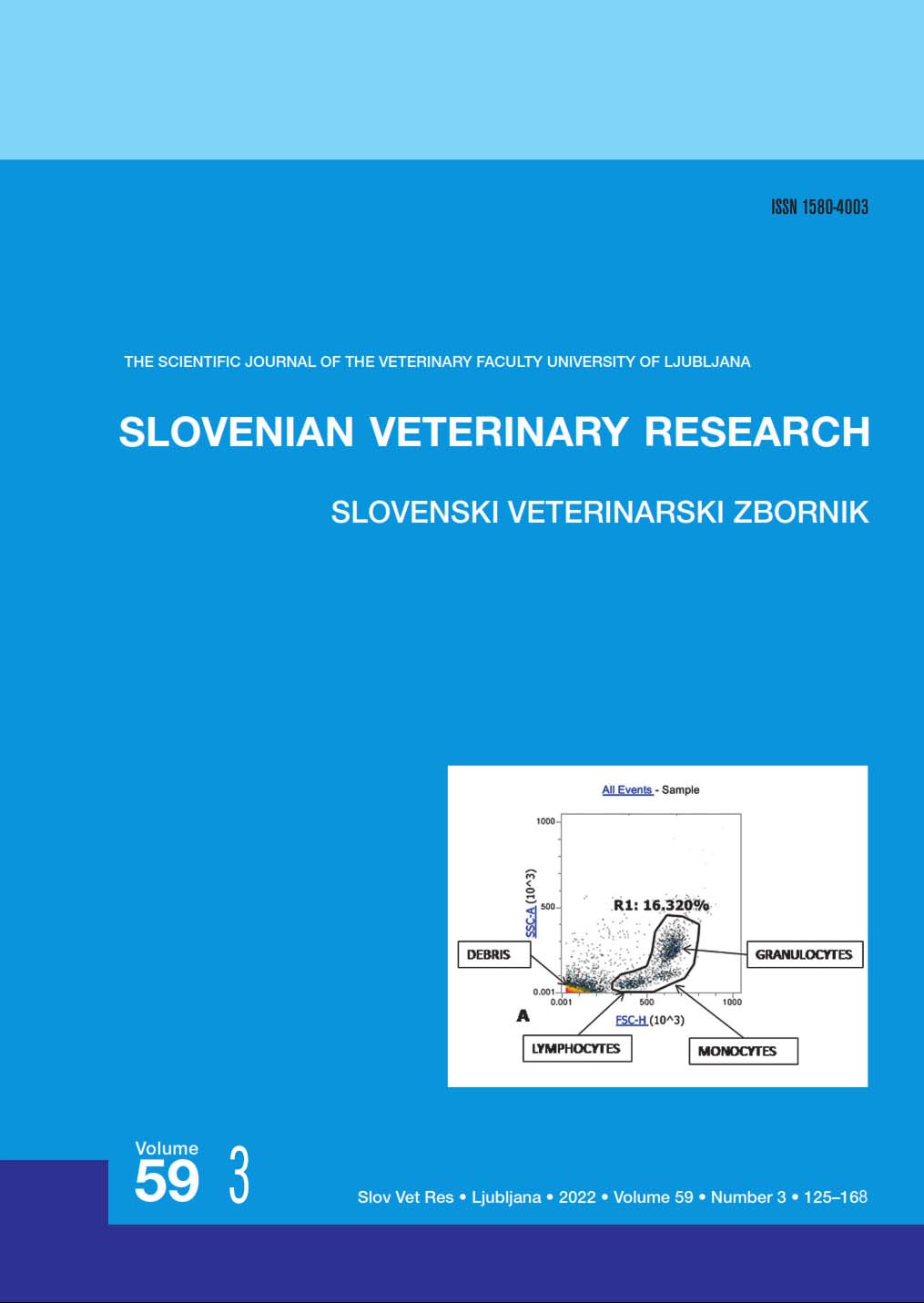
Lymphomas, leukemias and mast cell tumors belong to the most important group among all neoplasms affecting dog species. Diagnosis, staging and determining the cell type involved in a specific tumor represent a challenge for researchers and clinicians, and plays a crucial role in treatment efficacy and prognostic purposes. Many different gold standard techniques such as cytology, histopathology, immunohistochemistry and cytochemistry are used to routinely diagnose and stage these tumors. In the recent years flow cytometry is becoming more applicable in veterinary medicine since a wide number of health conditions can be analyzed in a short period of time with a high accuracy. Multiparametric analysis performed by flow cytometry is considered as one of the main advantages of this technique since cell populations can be analyzed for different superficial markers at the same time. Immunophenotyping and staging of tumor cell populations performed by flow cytometry can help in reaching a confirmatory diagnosis and appropriate prognosis of the disease. Moreover, many flow cytometric results have been linked to a high prognostic relevance especially in neoplastic disorders. However, flow cytometry results are compatible and should be interpreted in compliance with data obtained by histopathology, immunohistochemistry and cytology.
Key words: flow cytometry; antibodies; diagnosis; lymphoma; leukemia
UPORABNOST PRETOČNE CITOMETRIJE PRI PREPOZNAVANJU IN DOLOČANJU STADIJA LIMFOMA, LEVKEMIJE IN TUMORJEV MASTOCITOV PRI PSIH – PREGLED
Izvleček: Limfomi, levkemije in tumorji mastocitov so najpomembnejše skupine neoplazem, ki prizadenejo pse. Diagnostika, določanje stopenj tumorja in tipa celic v določenem tumorju predstavljajo izziv za raziskovalce in klinike in igrajo ključno vlogo pri učinkovitosti zdravljenja in postavljanju prognoze. Za rutinsko diagnosticiranje in določanje stopenj teh tumorjev se uporablja veliko različnih temeljnih metod, kot so citologija, histopatologija, imunohistokemija in citokemija. V zadnjih letih je pretočna citometrija vse bolj uporabljana metoda v veterinarski medicini, saj je mogoče v kratkem času in z visoko natančnostjo analizirati veliko število zdravstvenih stanj. Ena izmed najpomembnejših prednosti te tehnike je multiparametrična analiza, s katero lahko v populaciji celic istočasno analiziramo različne površinske označevalce. Določanje površinskih označevalcev in stopenj populacij tumorskih celic s pretočno citometrijo lahko pripomore k potrditvi diagnoze in postavitvi ustrezne prognoze bolezni. Številni rezultati pretočne citometrije so imeli pomemben prognostični pomen zlasti pri neoplastičnih obolenjih. Vendar je rezultate pretočne citometrije potrebno združiti in razlagati v skladu s podatki, pridobljenimi s histopatologijo, imunohistokemijo in citologijo.
Ključne besede: pretočna citometrija; protitelesa; diagnoza; limfom; levkemija
References
● 1. Prier JE, Brodey RS. Canine neoplasia. A proto-type for human cancer study. Bull WHO 1963; 29: 331–44.
● 2. Fournel-Fleury C, Magnol JP, Bricaire P, et al. Cy-tohistological and immunological classification of canine malignant lymphomas: comparison with human non-Hodgkin's lymphomas. J Comp Pathol 1997; 117(1): 35–59.
● 3. Ponce F, Marchal T, Magnol JP, et al. A morpho-logical study of 608 cases of canine malignant lymphoma in France with a focus on comparative similarities be-tween canine and human lymphoma morphology. Vet Pathol 2010: 47: 414–33.
● 4. Valli EV, San Myint M, Barthel A, et al. Classifica-tion of canine malignant lymphomas according to the World Health Organization criteria. Vet Pathol 2011: 48 (1): 198–211.
● 5. Morse III CH, Anver RM, Torgny N, et al. Be-thesda proposals for classification of lymphoid neo-plasms in mice. Blood 2002; 100: 246–58.
● 6. Kogan CS, Ward MJ, Anver RM, et al. Bethesda proposals for classification if nonlymphoid hematopoiet-ic neoplasms in mice. Blood 2002; 100: 238–45.
● 7. Garnica KT, Lesbon CCJ, Avila MCFCA, et al. Liquid biopsy based on small extracellular vesicles pre-dicts chemotherapy response of canine multicentric lymphomas. Sci Rep 2020; 10(1): e20371. doi: 10.1038/s41598-020-77366-7.
● 8. Rehg EJ, Bush D, Ward MJ. The utility of im-munohistochemistry for the identification of hemato-poietic and lymphoid cells in normal tissues and inter-pretation of proliferative and inflamma- tory lesions of mice and rats. Toxicol Pathol 2012; 40: 345–74.
● 9. Stokol T, Schaefer DM, Shuman M, Belcher N, Dong L. Alkaline phosphatase is a useful cytochemical marker for the diagnosis of acute myelomonocytic and monocytic leukemia in dogs. Vet Clin Pathol 2015; 44 (1): 79–93.
● 10. Swerdlow SH, Campo E, Harris NL, et al. WHO Classification of tumours. 4th ed. Volume 2. Geneva : World Health Organization, 2008.
● 11. Rout DE, Labadie DJ, Yoshimoto AJ, et al. Clini-cal outcome and prognostic factors in dogs with B-cell chronic lymphocytic leukemia: a retrospective study. J Vet Intern Med 2021; 35: 1918–28.
● 12. Misdrop W. Mast cells and canine mast cell tu-mours. Vet Q 2004; 26: 156–69.
● 13. Blackwood L, Murphy S, Buracco P, et al. Euro-pean consensus document on mast cell tumors in dogs and cats. Vet Comp Oncol 2012; 10: 1–29.
● 14. Kiupel M, Webster JD, Bailey KL, et al. Proposal of a 2-tier histologic grading system for canine cutane-ous mast cell tumors to more accurately predict biologi-cal behavior. Vet Pathol 2011; 48: 147–55.
● 15. Patnaik AK, Ehler WJ, Mac Ewen EG. Canine cutaneous mast cell tumour: morphologic grading and survival time in 83 dogs. Vet Pathol 1984; 21: 469–74.
● 16. Kiupel M, Webster JD, Kaneene JB, Miller R, Yuzbasivan-Gurkan V. The use of KIT and tryptase expression patterns as prognostic tools for canine cuta-neous mast cell tumors. Vet Pathol 2004; 41: 371–7.
● 17. Scase TJ, Edwards D, Miller J, et al. Canine mast cell tumors: correlation of apoptosis and proliferation markers with prognosis. J Vet Intern Med 2006; 20: 151–8.
● 18. Krick EL, Billings AP, Shofer FS, Watanabe S, Sorenmo KU. Cytological lymph node evaluation in dogs with mast cell tumours: association with grade and survival. Vet Comp Oncol 2009; 7(2): 130–8.
● 19. Garrett DL. Canine mast cell tumors: diagnosis, treatment and prognosis. Vet Med Res Rep 2014; 5: 49–58.
● 20. Adan A, Alizazada G, Kiraz Y, Baran Y, Nalbant A. Flow cytometry: basic principles and applications. Crit Rev Biotechnol 2017; 37(2): 163–76.
● 21. Aebishera D, Bartusikb D, Tabarkiewiczc J. Laser flow cytometry as a tool for the advancement of clinical medicine. Biomed Pharmacother 2017; 85: 434–43.
● 22. McKinnon MK. Flow cytometry: an overview. Curr Protoc Immunol 2019; 120: 5.1.1–5.1.11. doi: 10.1002/cpim.40
● 23. Mckinnon MK. Multiparameter conventional flow cytometry. Methods Mol Biol 2018; 1678: 139–50.
● 24. Miniscalco B, Poggi A, Martini V, et al. Flow cy-tometric characterization of S-phase fraction and ploidy in lymph node aspirates from dogs with lymphoma. J Comp Pathol 2018; 161: 34–43.
● 25. Sulce M, Marconato L, Martano M, et al. Utility of flow cytometry in canine primary cutaneous and matched nodal mast cell tumor. Vet J 2018; 242: 15–23.
● 26. Martini V, Melega M, Riondato F, et al. A retro-spective study of flow cytometric characterization of suspected extranodal lymphomas in dogs. J Vet Diagn Invest 2018; 30(6): 830–6.
● 27. Riondato F, Martini V, Poggi A, et al. Identifica-tion of a suitable internal control for fluorescence analy-sis on canine peripheral blood samples. Vet Immunol Immunopathol 2016; 172: 38–42.
● 28. Poggi, A, Miniscalco, B, Morello, E, et al. Flow cytometric evaluation of ki67 for the determination of malignancy grade in canine lymphoma. Vet Comp Oncol 2013; 13(4): 475–80.
● 29. Comazzi S, Gelain ME, Martini V, et al. Im-munophenotype predicts survival time in dogs with chronic lymphocytoc leukemia. J Vet Intern Med 2011; 25(1): 100–6.
● 30. Menon V, Thomas R, Ghale RA, Reinhard C, Jan Pruzak. Flow cytometry protocols for surface and intra-cellular antigen analyses of neural cell types. J Vis Exp 2014; 94: e52241. doi: 10.3791/52241.
● 31. Park Y, Abihssira-Garcia SI, Thalmann S, et al. Imaging flow cytometry protocols for examining phago-cytosis of microplastics and bioparticles by immune cells of aquatic animals. Front Immunol 2020; 11: e203. doi: 10.3389/fimmu.2020.00203.
● 32. Seelig DM, Avery P, Webb T, et al. Canine T-zone lymphoma: unique immunophenotypic features, outcome, and population characteristics. J Vet Intern Med 2014; 28(3): 878–86.
● 33. Baumgarth N, Roederer M. A practical approach to multicolor flow cytometry for immunophenotyping. J Immunol Methods 2000; 243: 77–97.
● 34. Riondato F, Comazzi S. Flow cytometry in the diagnosis of canine B-cell lymphoma. Front Vet Sci 2021; 8: e600986. doi: 10.3389/ fvets.2021.600986.
● 35. Comazzi S, Riondato F. Flow Cytometry in the diagnosis of canine T-cell lymphoma. Front Vet Sci 2021; 8: e600963. doi: 10.3389/fvets.2021.600963
● 36. Stokol T, Nickerson AG, Shuman M, Belcher N. Dogs with acute myeloid leukemia have clonal rear-rangements in T and B cell receptors. Front Vet Sci 2017; 4: e76. doi: 10.3389/ fvets.2017.00076
● 37. Comazzi S, Avery PR, Gardern OA, Rindato F, Rutgen B, Vernau W. European canine lym- phoma net-work consensus recommendations for reporting flow cytometry in canine hematopoietic neoplasms. Cytome-try B Clin Cytom 2017; 92(5): 411–9.
● 38. Drescher H, Weiskirchen S, Weiskirchen R. Flow cytometry: a blessing and a curse. Biomedicines 2021; 9(11): e1613. doi: 10.3390/biomedicines9111613
● 39. Janke JL, Mullighan GC, Dang J, Rehg EJ. Im-munophenotyping of murine percursor B-cell leuke-mia/lymphoma: a comparison of immunohistochemistry and flow cytometry. Vet Pathol 2019; 56: 950–8.
Downloads
Published
How to Cite
Issue
Section
License
Copyright (c) 2022 Majlind Sulçe, Albana Munga, Doriana Beqiraj, Enkeleda Ozuni, Pëllumb Zalla, Gerald Muca, Xhelil Koleci

This work is licensed under a Creative Commons Attribution-ShareAlike 4.0 International License.
