
COMPARATIVE EVALUATION OF THE KOMODO DRAGON (Varanus komodoensis) AND THE GREEN IGUANA (Iguana iguana) SKULL BY THREE DIMENSIONAL COMPUTED TOMOGRAPHIC RECONSTRUCTION
DOI:
https://doi.org/10.26873/SVR-1330-2021Abstract
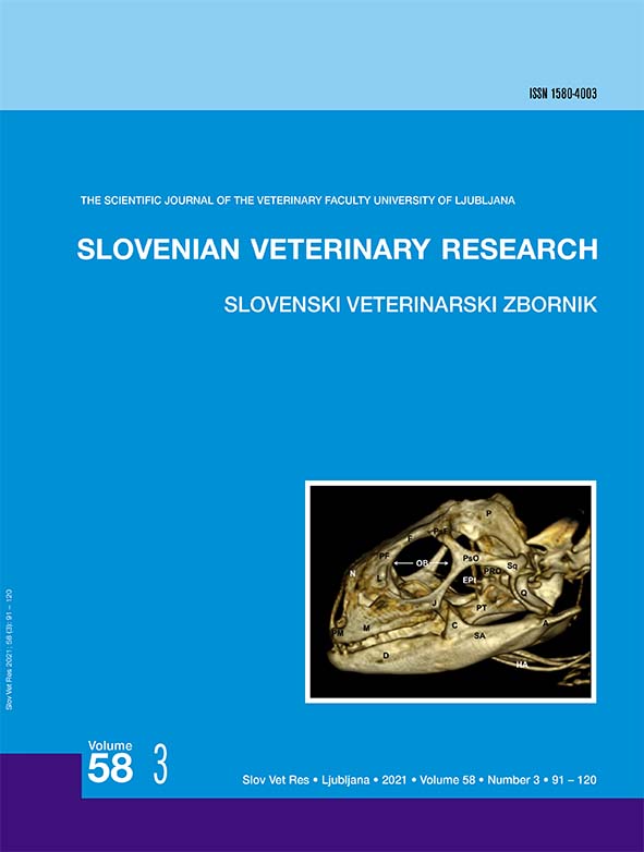
The purpose of this paper was to do a comparative evaluation of the skull of two species of lizards, the Komodo dragon (Varanus komodoensis) and the Green Iguana (Iguana iguana), by three-dimensional computed tomographic reconstruction. Images provided by this method give excellent anatomic detail of the skull. Therefore, essential differences in the configuration of the orbit and the lateral bones of the neurocranium were visualized in lateral and dorsal reconstructed images. The images obtained by tridimensional computed tomographic reconstruction can be a valuable diagnostic aid for the clinical evaluation of several head disturbances in lizards.
Key words: computed tomography; 3D reconstruction; anatomy; skull; lizards
PRIMERJAVA LOBANJ KOMODOŠKEGA VARANA (Varanus komodoensis) IN ZELENEGA LEGVANA (Iguana iguana) S POMOČJO TRIDIMENZIONALNE RAČUNALNIŠKE TOMOGRAFSKE REKONSTRUKCIJE
Izvleček: Namen prispevka je bil s tridimenzionalno računalniško tomografsko rekonstrukcijo opraviti primerjalno oceno lobanje dveh vrst kuščarjev, komodoškega varana (Varanus komodoensis) in zelenega legvana (Iguana iguana). Slike, pridobljene s to metodo, prikažejo odlične anatomske podrobnosti lobanje. Zato so bile na stranskih in dorzalnih rekonstrukcijah slik vidne bistvene razlike v zgradbi orbitalnega področja in stranskih kosti nevrokranija med obema vrstama kuščarjev. Slike, pridobljene s tridimenzionalno računalniško tomografsko rekonstrukcijo, so lahko dragocena diagnostična pomoč pri klinični oceni večih napak glave pri kuščarjih.
Ključne besede: računalniška tomografija; 3D rekonstrukcija; anatomija; lobanja; kuščarji
References
(1.) Gumpenberger M. Diagnostic imaging of reproductive tract disorders in reptiles. Vet Clin North Am Exot Anim Pract 2017; 20(2): 327–43.
(2.) Hernández C, Peloso PL, Bolívar W, Daza JD. Skull morphology of the lizard Ptychoglossus vallensis (Squamata:Alopoglossidae) with comments on the variation within Gymnophthalmoidea. Anat Rec 2019; 302(7): 1074–92.
(3.) Banzato T, Hellebuyck T, Van Caelenberg A, Saunders JH, Zotti A. A review of diagnostic imaging of snakes and lizards. Vet Rec 2013; 173(2): 439.
(4.) Banzato T, Russo E, Di Toma A, Palmisano G, Zotti A. Anatomic imaging of the Boa constrictor head: a comparison between radiography, computed tomography and cadaver anatomy. Am J Vet Res 2011; 72(12): 1592–9.
(5.) Banzato T, Selleri P, Veladiano IA, Martin A, Zanetti E, Zotti A. Comparative evaluation of the cadaveric, radiographic and computed tomographic anatomy of the heads of green iguana (Iguana iguana), common tegu (Tupinambis merianae) and bearded dragon (Pogona vitticeps). BMC Vet Res 2012; 8: e53. doi: 10.1186/1746-6148-8-53
(6.) Lauridsen H, Hansen K, Wang T, et al. Inside out: modern imaging techniques to reveal animal anatomy. PLoS ONE 2011; 6(3): e17879. doi: 10.1371/journal.pone.0017879
(7.) Wilken AT, Middleton KM, Sellers KC, Cost IN, Holliday CM. The roles of joint tissues and jaw muscles in palatal biomechanics of the savannah monitor (Varanus exanthematicus) and their significance for cranial kinesis. J Exp Biol 2019; 222 (Pt 18): jeb201459.
(8.) Holliday CM, Tsai HP, Skiljan RJ, George ID, Pathan S. A 3D iInteractive model and atlas of the jaw musculature of Alligator mississippiensis. PLoS One 2013; 8(6): e62806. doi: 10.1371/journal.pone.0062806
(9.) Arencibia A, Melian A, Oros J. Anatomic interactive atlas of the loggerhead sea turtle (Caretta caretta) head. Animals 2021; 11(1): e198. doi: 10.3390/ani11010198
(10.) Knipe FM. Principles of neurological imaging of exotic animal species. Vet Clin North Am Exot Anim Pract 2007; 10(3): 893–907.
(11.) Krautwald-Junghanns ME, Pees M, Reese S. Diagnostic imaging of exotic pets. Hannover : Schlutersche, 2011: 310–459.
(12.) Arencibia A, Corbera JA, Ramírez G, et al. Anatomical assessment of the thorax in the neonatal foal using computed tomography angiography, sectional anatomy and gross dissections. Animals 2020; 10(6): e1045. doi: 10.3390/ani10061045
(13.) Zafra R, Carrascosa C, Suarez-Bonnet A, et al. Three-dimensional reconstruction by computed tomography of an undifferentiated sarcoma in a dog. J Appl Anim Res 2012; 40(4): 289–91.
(14.) Jaber JR, Carrascosa C, Arencibia A, Corbera JA, Ramirez AS, Melian C. 3-D computed tomography reconstruction: another tool to teach anatomy in the veterinary colleges. Iran J Vet Res 2018; 19(1): 1-2.
(15.) Daza JD, Bauer AM. The circumorbital bones of the Gekkota (Reptilia: Squamata). Anat Rec 2010; 293(3): 402–13.
(16.) Moselhy AA, Mahdy, E. Comparative three dimensionalcomputed tomography (CT) scans and anatomical investigation of rabbit (Oryctolagus cuniculus) and cat (Felis domestica) skull. Slov Vet Res 2019; 56 (Suppl 22): 365–79.
Downloads
Additional Files
Published
How to Cite
Issue
Section
License
Copyright (c) 2021 Sara Pérez, Mario Encinoso, Manuel Morales, Alberto Arencibia, Alejandro Suárez-Bonnet, Eligia González-Rodríguez, Jose Raduan Jaber

This work is licensed under a Creative Commons Attribution-ShareAlike 4.0 International License.
